
Views: 0 Author: Site Editor Publish Time: 2026-08-15 Origin: Site


In analytical chemistry and molecular biology, spectrophotometric data integrity relies entirely on the optical neutrality of the sample container. Minor material interference invalidates quantitative analysis immediately. Laboratories frequently face baseline drift, signal attenuation, and irreproducible results when measuring in the ultraviolet spectrum below 340 nm. This failure occurs due to the inherent UV-absorbing properties of standard optical glass and disposable plastics.
To achieve precise, reproducible transmission in the UV range, laboratory managers must standardize their instrumentation with materials engineered for broad-spectrum optical clarity. This technical evaluation examines the specifications, operational trade-offs, and return on investment of utilizing a quartz cuvette for high-stakes analytical workflows. We will analyze material limitations, configuration selection, and the implementation of strict handling protocols to prevent cross-contamination and optical degradation in your facility.
Unrestricted UV Transmission: Quartz provides an extended optical transmission range from 190 nm to 2,500 nm, preventing the signal blockage that occurs with glass or plastic below 340 nm.
Chemical and Thermal Resilience: Fused silica quartz withstands aggressive organic solvents, strong acids, and extreme temperature fluctuations without degrading or leaching.
Pathlength Precision: High-quality quartz cuvettes are manufactured to strict tolerances, ensuring exact pathlengths (typically 10 mm) critical for accurate Beer-Lambert Law calculations.
Long-Term ROI: While the initial capital expenditure is higher, the reusability, durability, and prevention of false readings make quartz the most cost-effective choice for rigorous, high-volume UV-Vis analysis.
Standard optical glass and polystyrene or polymethyl methacrylate (PMMA) plastics possess molecular structures that actively absorb ultraviolet radiation. When light passes through these materials at wavelengths below 340 nm, the material itself acts as a physical barrier. The aromatic rings in polystyrene, for example, exhibit strong pi-electron transitions that absorb heavily around 260 nm and 280 nm. This absorption blocks the light source from reaching the detector.
Consequently, the spectrophotometer records false absorbance peaks that belong to the container rather than the sample. This interference makes accurate quantification of nucleic acids or proteins impossible using standard materials. If an operator attempts to blank the instrument using a plastic cell at 260 nm, the photodetector receives almost zero transmitted light. The instrument will either throw an error or amplify the detector gain to maximum, resulting in massive baseline noise that drowns out the actual sample signal.
A successful sample cell must remain optically invisible to the spectrometer across the entire target wavelength range. The material purity of a UV spectrophotometer cuvette directly dictates baseline noise and the overall signal-to-noise ratio. If the cell walls absorb even a fraction of the incident light, baseline drift occurs as the lamp intensity fluctuates or the material degrades under prolonged UV exposure.
High-purity silica ensures that the detector only measures the absorbance of the analyte. Maintaining a stable baseline guarantees reproducible data across multiple testing cycles. In high-throughput environments, baseline stability prevents the need for constant re-blanking. Operators can run dozens of samples sequentially with confidence that the optical path remains consistent. Any deviation in the baseline directly translates to concentration errors, which is unacceptable in clinical diagnostics or pharmaceutical quality control.
Fused silica offers an exceptional optical transmission curve. It remains highly transparent from the deep UV region at 190 nm through the visible spectrum and into the near-infrared at 2500 nm. This broad utility allows laboratories to use a single cell type for diverse applications. You can perform nucleic acid quantification at 260 nm, measure protein concentrations at 280 nm, and conduct standard colorimetric assays in the visible range without changing the sample container.
This wide transmission window eliminates the logistical headache of swapping cell types between different assays. When a lab standardizes on fused silica, operators do not need to worry about accidentally using a visible-only glass cell for a UV assay. The transmission efficiency of high-grade silica typically exceeds 80% across this entire range, with the remaining 20% lost primarily to surface reflection rather than internal absorption. This high transmission ensures maximum light reaches the detector, improving the dynamic range of the instrument.
Understanding material classifications prevents analytical errors. Synthetic Fused Silica is optimized for Far-UV applications. Infrared Quartz (Infrasil) features ultra-low hydroxyl (OH) content, preventing water absorption bands from interfering with measurements in the near-infrared spectrum. Optical glass is restricted entirely to the visible range.
| Material Type | Optimal Wavelength Range | Hydroxyl (OH) Content | Common Analytical Applications |
|---|---|---|---|
| Optical Glass | 340 nm – 2,500 nm | High | Routine visible range colorimetry, cell density (OD600), Bradford assays. |
| Synthetic Fused Silica (UV) | 190 nm – 2,500 nm | High (1000+ ppm) | DNA/RNA quantification, protein assays, deep UV scanning, HPLC detectors. |
| Infrared Quartz (Infrasil) | 220 nm – 3,500 nm | Ultra-Low (<8 ppm) | Near-infrared spectroscopy, moisture analysis, specialized IR scanning. |
Circular Dichroism (CD) spectroscopy is a highly sensitive technique requiring absolute polarization neutrality. Plastics, standard glass, and poorly annealed silica often contain internal stresses from the manufacturing process. These stresses cause birefringence, which distorts circularly polarized light. When the light polarization is altered by the container, the resulting CD spectrum is corrupted.
Using strain-free silica ensures that the polarization state of the light remains unaltered as it passes through the cell walls. Manufacturers achieve this through specialized annealing processes that slowly cool the material, relieving all internal mechanical stress. This neutrality is mandatory for valid CD measurements and accurate protein secondary structure analysis. Without strain-free windows, differentiating between alpha-helices and beta-sheets becomes impossible due to the high background noise generated by the cell itself.
Analytical workflows frequently utilize aggressive reagents. Concentrated acids, strong bases, and organic solvents like acetone, acetonitrile, or chloroform will rapidly melt, cloud, or degrade plastic alternatives. Fused silica demonstrates superior chemical inertness. It does not leach plasticizers, mold release agents, or unreacted monomers into the sample.
Leaching is a severe problem in trace-level UV analysis because many plasticizers absorb strongly in the UV range. If a solvent extracts these compounds from a plastic cell, the sample absorbance will artificially inflate. Fused silica's robust solvent compatibility ensures the cell remains optically clear and structurally intact over thousands of testing cycles. The only chemicals that significantly attack fused silica are hydrofluoric acid (HF) and hot, highly concentrated alkalis.
Silica possesses an exceptionally low coefficient of thermal expansion (approximately 0.55 x 10^-6 /°C). This thermal stability is vital for thermodynamic studies, enzyme kinetics, and DNA melting point analyses. In these applications, the sample cell undergoes rapid heating and cooling cycles via thermoelectric Peltier systems built into the spectrophotometer cell holder.
Unlike plastics or standard glass, which may warp, change pathlength, or crack under thermal stress, fused silica maintains its precise dimensions. When conducting a DNA melt curve analysis from 25°C to 95°C, the pathlength must remain exactly 10 mm. If the material expands, the pathlength increases, artificially altering the absorbance readings and skewing the calculated melting temperature (Tm). Fused silica prevents this thermal distortion entirely.
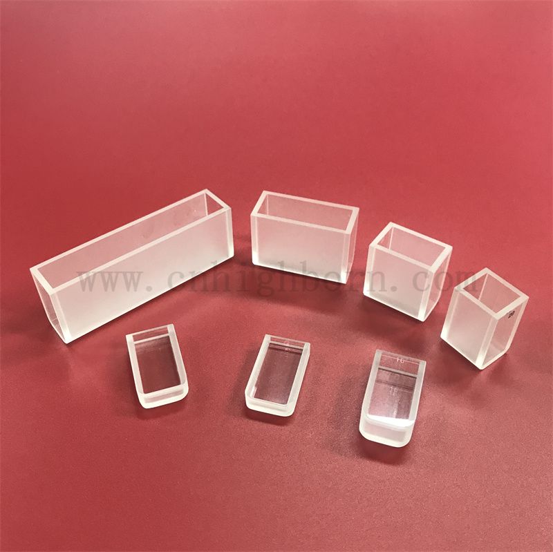
The standard two side clear silica cuvette is the primary tool for absorption and transmission spectrophotometry. It features two polished optical windows for the light path and two frosted or blackened sides. The frosted sides prevent internal light scattering and unwanted reflections that can bounce back into the detector and cause non-linear absorbance responses at high concentrations.
These frosted sides also provide a designated handling surface. Operators must grip the cell exclusively by these non-optical faces. This design ensures that users do not leave fingerprints, skin oils, or glove residues on the critical optical windows during sample loading. Even a microscopic smudge on the optical face will scatter the UV beam, leading to immediate baseline elevation and inaccurate concentration calculations.
Fluorescence spectroscopy requires a different optical geometry. Because fluorescence emission is measured at a 90-degree angle to the excitation light source, the cell must have four optically clear sides. Using a standard two-sided cell for fluorescence will block the emitted light from reaching the detector, resulting in a zero reading.
Four-sided clear configurations are also necessary for dynamic light scattering (DLS) and nephelometry applications. When selecting a four-sided cell, operators must be exceptionally careful during handling, as there are no frosted sides to grip. Specialized cell lifters or handling strictly by the extreme top edge is required to maintain optical clarity on all four transmission faces.
Sample volume constraints dictate cell geometry. Standard macro configurations typically require 3.5 mL of sample to ensure the light beam passes entirely through the liquid and does not strike the meniscus. When dealing with precious or low-yield biological samples, laboratories transition to semi-micro or micro-cuvettes.
Semi-Micro Cells: Typically require 1.0 mL to 1.5 mL of sample. The internal width is reduced, but the 10 mm pathlength remains constant.
Micro Cells: Can analyze volumes as low as 50 µL. These feature black masking around the small optical window to prevent the light beam from passing through the empty space above the liquid.
Flow-Through Cells: Integrate directly into automated liquid handling systems, dissolution testers, or chromatography detectors. They allow for continuous inline monitoring without manual sample transfer.
When using micro cells, operators must verify the Z-dimension (beam height) of their specific spectrophotometer. Instruments typically have a Z-dimension of either 8.5 mm or 15 mm. If a micro cell with an 8.5 mm window is placed in a 15 mm instrument, the light beam will strike the black masking instead of the sample.
High-end UV-transparent plastics attempt to bridge the gap between disposable convenience and UV functionality. However, they consistently exhibit baseline variability, higher background noise, and sharp transmission drop-offs below 220 nm. Batch-produced disposable plastics carry inherent risks of optical inconsistencies, molding defects, and pathlength variations from one cell to the next.
Fused silica eliminates these variables. It delivers unmatched optical clarity and manufacturing tolerances, ensuring that analytical success criteria are met without the risk of container-induced data artifacts. When measuring highly concentrated samples where the absorbance approaches 2.0 or 3.0, the stray light performance of the cell becomes critical. Plastics scatter too much light to be reliable at high optical densities, whereas polished silica maintains strict linearity.
Evaluating the financial impact requires looking beyond the initial purchase price. Single-use plastics demand continuous procurement, dedicated storage space, and ongoing biohazard disposal costs. A high-volume laboratory running hundreds of assays weekly will quickly exceed the cost of a premium silica cell through plastic consumable spending alone.
The true cost-efficiency of silica lies in failure prevention. If a plastic cell causes a false reading during a critical pharmaceutical batch release, the cost of the investigation, sample re-runs, and potential batch quarantine dwarfs the price of optical equipment. By standardizing on reusable silica, laboratories eliminate the consumable variable from their root-cause analysis when troubleshooting unexpected assay results.
The analytical market contains poorly manufactured alternatives that compromise data integrity. You must verify manufacturer tolerances before implementation. A strict pathlength accuracy of ±0.01 mm is necessary to adhere to the Beer-Lambert Law. If the pathlength is 10.1 mm instead of 10.0 mm, every concentration calculation will be off by 1%.
Furthermore, window parallelism must be exact. Non-parallel windows act as a prism, deflecting the light beam away from the detector. This beam deflection causes severe measurement errors, particularly in instruments with small detector apertures. High-quality cells are assembled using direct fusion techniques rather than adhesives. Adhesives can dissolve in organic solvents, leading to cell failure and sample leakage inside the instrument compartment.
Improper cleaning introduces trace organic residues or protein carryover, which absorb heavily in the UV range. Standardize your cleaning protocols using specialized washers and detergents. Avoid highly alkaline glass cleaners, which will etch the silica surface over time, permanently clouding the optical windows.
Immediately after use, rinse the cell with the appropriate solvent to remove the bulk of the sample.
Soak in a specialized optical detergent or a mild nitric acid solution (e.g., 3M HNO3) to break down protein residues.
Rinse thoroughly with copious amounts of ultrapure deionized water (18.2 MΩ·cm).
Perform a final rinse with analytical-grade ethanol or methanol to facilitate rapid drying.
Dry using a stream of clean, oil-free nitrogen gas or compressed air. Never air-dry, as this leaves water spots on the optical faces.
Physical damage permanently degrades optical performance. Operators must only handle the cell by the frosted sides. Never wipe optical windows with standard paper towels, laboratory wipes, or clothing. These materials contain abrasive fibers that will create micro-scratches on the polished silica surface. Use only lint-free photographic lens paper if wiping is absolutely necessary.
Store the cells in dedicated silicone, PTFE, or soft plastic racks. Never store them loose in a drawer or in hard metal racks where they can chip. When inserting the cell into the spectrophotometer, ensure the optical faces are aligned with the light beam and push down gently. Forcing a cell into a tight holder can cause micro-fractures along the fused seams.
Audit your current laboratory standard operating procedures to identify all assays requiring measurements below 340 nm or involving aggressive organic solvents.
Verify the pathlength tolerances, Z-dimension requirements, and material purity grades with your manufacturer before procuring new optical cells.
Implement strict, standardized cleaning and handling protocols to prevent micro-scratches, protein carryover, and surface etching.
Transition away from disposable plastics for any high-precision quantitative analysis to eliminate baseline variability and molding defects.
A: It typically ranges from 190 nm to 2,500 nm. This covers the deep UV, visible, and near-infrared spectrums, providing broad utility unlike standard glass which cuts off around 340 nm.
A: Beyond manufacturer labeling, fused silica will transmit UV light (such as at 260 nm) without absorbing it. Glass will block this transmission completely. UV transmission remains the definitive functional test.
A: Standard plastics like polystyrene absorb UV light and cannot be used. Specialized UV-transparent plastics work down to approximately 230 nm, but they lack the chemical resistance and strict pathlength tolerances of fused silica.
A: Handle it only by the frosted sides to keep fingerprints off the optical path. Clean with specialized detergents, ethanol, or mild acid solutions. Never use abrasive brushes or highly alkaline solutions that etch silica.
A: The cost is driven by the high-purity raw material, the extreme melting temperatures required for manufacturing, and the rigorous optical polishing needed to guarantee exact pathlengths and parallel optical surfaces.
A: The industry standard pathlength is 10 mm. This simplifies calculations for the Beer-Lambert Law. However, pathlengths ranging from 1 mm to 100 mm are available for specific concentration requirements.
A: CD measurements require specialized strain-free quartz. Standard materials can possess internal stresses that cause birefringence, which distorts the polarization of light and ruins the sensitive CD signal.